How a Phone Camera Measures Your Breathing Rate
Discover the science behind contactless vitals. Learn how optical flow and rPPG allow a standard smartphone camera to accurately measure patient respiratory rate.
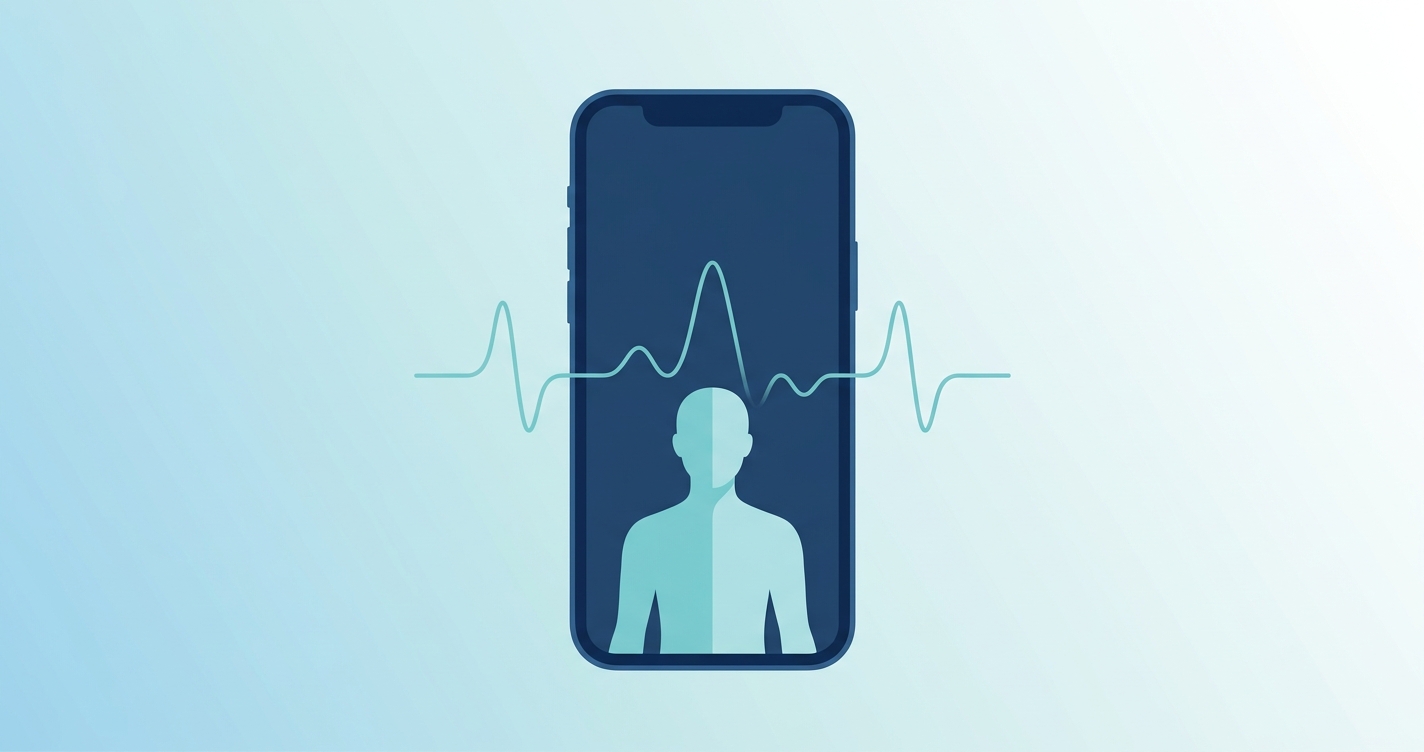
The widespread adoption of telehealth has resolved the challenge of connecting patients and providers over video, but clinical informatics teams still face a barrier: acquiring objective physiological data without shipping hardware to the patient's home. Respiratory rate is a critical indicator of clinical deterioration, yet it is notoriously difficult to assess accurately through a standard video feed. Fortunately, software advancements now allow standard smartphone sensors to perform clinical-grade assessments. Understanding how a camera measures breathing rate is essential for health systems evaluating virtual visit vitals capture platforms. By analyzing subtle micro-color changes in the skin and tracking microscopic physical movements, standard device optics can provide real-time respiratory metrics, bridging the gap between an in-person exam and a remote consultation.
"Recent studies on contactless vital sign capture report a mean absolute error of just 0.5 breaths per minute compared to standard capnography, demonstrating that optical flow and photoplethysmography algorithms can achieve clinical viability without patient wearables." - 2022 Systematic Review of Contactless Respiratory Rate Monitoring
How a camera measures breathing rate: physiology and physics
When a patient asks how a camera measures breathing rate, the answer lies in two parallel computational techniques: remote photoplethysmography (rPPG) and optical flow analysis. These technologies work together to turn a commercial smartphone into a remote clinical sensor.
Remote photoplethysmography operates on the principle that human skin changes color slightly with every heartbeat. As the heart pumps, blood volume in the microvascular bed of the facial skin increases and decreases. Blood absorbs ambient light, particularly in the green spectrum. When blood volume is high during systole, more light is absorbed and less reflects back to the camera sensor. When blood volume is low during diastole, more light is reflected. High-resolution cameras on modern smartphones capture these minute fluctuations in red, green, and blue pixel intensities.
While rPPG is primarily known for extracting heart rate, it is equally effective at determining telehealth respiratory rate. This is due to a physiological phenomenon known as respiratory sinus arrhythmia. As a person inhales, their heart rate slightly increases; as they exhale, their heart rate decreases. Furthermore, the physical mechanics of breathing cause baseline shifts in the blood volume and alter the amplitude of the pulse signal. Advanced algorithms isolate these secondary respiratory frequencies from the primary cardiac signal to calculate breaths per minute.
Simultaneously, optical flow analysis monitors the physical mechanics of respiration. Optical flow algorithms track the movement of specific pixels over time. When applied to a video feed of a patient, the software identifies regions of interest, such as the shoulders, chest, and abdomen. The system measures the rhythmic rise and fall of these regions. By fusing the micro-color data from rPPG with the mechanical movement data from optical flow, clinical informatics teams can capture contactless vitals with high reliability.
A critical consideration in optical health measurement is skin tone inclusivity. Because melanin absorbs light, darker skin tones reflect less light back to the camera sensor, which can reduce the signal-to-noise ratio in rPPG applications. To address this, modern clinical algorithms do not rely on a single color channel. Instead, they dynamically weigh the red, green, and blue signals based on the individual patient's baseline skin tone and ambient lighting conditions. By processing the full spectrum of available light and corroborating the data with mechanical motion tracking, the software ensures equitable accuracy across all patient demographics.
Comparing modalities: camera-based vs. contact sensors
Health systems evaluating remote monitoring solutions must compare software-based tools against traditional hardware. The table below outlines how camera-based systems stack up against standard contact sensors.
| Feature | Camera-Based Measurement | Wearable Chest Strap | Manual Clinical Observation | | :--- | :--- | :--- | :--- | | Data Capture Method | Optical flow and rPPG | Strain gauges or ECG | Visual counting by provider | | Hardware Required | Patient smartphone | Shipped proprietary device | None (requires live provider) | | Patient Burden | Zero contact | Requires wearing device | Requires sitting still for 60 seconds | | Scalability | Immediate across populations | Limited by supply chain | Limited by provider time | | Typical Accuracy (MAE) | 0.5 to 1.0 breaths/min | 0.5 breaths/min | Variable (prone to human error) |
Core technical steps to check breathing rate with phone
Transforming raw video data into a clinically actionable vital sign requires a sophisticated computational pipeline. The process to check breathing rate with phone cameras generally follows four distinct stages:
- Region of Interest Selection: The software uses facial detection and body pose estimation models to identify the optimal areas for measurement. For rPPG, this is typically the forehead and cheeks, where the microvascular bed is dense and visible. For optical flow, the software targets the upper chest and shoulders.
- Signal Extraction: The camera captures raw pixel data at 30 to 60 frames per second. The algorithm extracts the average color intensities from the facial regions and the vertical displacement coordinates from the chest regions.
- Filtering and Amplification: The raw signals contain noise from ambient light changes, minor head movements, and camera auto-focus adjustments. Bandpass filters isolate frequencies between 0.15 Hz and 0.5 Hz, which corresponds to the typical human respiratory rate of 9 to 30 breaths per minute.
- Respiratory Rate Calculation: The filtered signals undergo spectral analysis, often using a Fast Fourier Transform, to identify the dominant frequency. This frequency is multiplied by 60 to calculate the final breaths per minute.
Industry applications in health systems
The ability to capture virtual visit vitals without deploying hardware fundamentally changes the economics and clinical depth of telehealth programs.
Chronic care management
Patients with congestive heart failure, chronic obstructive pulmonary disease, and asthma require frequent monitoring. An elevated respiratory rate is often the earliest leading indicator of a pulmonary exacerbation. By checking a patient's breathing rate through a routine video check-in, care teams can intervene days before the patient requires emergency department admission.
Triage and emergency room diversion
Health systems are increasingly launching virtual triage and urgent care services. When a patient reports a fever and cough, the provider needs objective data to differentiate between a mild viral infection and severe respiratory distress. Contactless vitals provide the objective data needed to confidently manage the patient at home or direct them to an emergency facility.
Post-Discharge Monitoring
The transition from the hospital to the home is a vulnerable period. Supplying every discharged patient with a pulse oximeter or respiratory strap is logistically and financially unfeasible for most health systems. Camera-based measurement allows care teams to monitor respiratory trends using the hardware the patient already owns.
The success of post-discharge monitoring relies heavily on seamless data integration. When a patient completes a virtual check-in, the captured respiratory rate and heart rate data must flow directly into the electronic health record (EHR). This automated documentation prevents data silos and ensures the patient's primary care provider and specialist have immediate access to the trends. By standardizing this data flow, health systems can trigger automated clinical alerts if a patient's breathing rate exceeds safe thresholds, enabling proactive outreach before an emergency occurs.
Current research and evidence
The transition of rPPG and optical flow from computer science labs to clinical applications is documented by rigorous peer-reviewed research. A foundational study on robust respiration detection from remote photoplethysmography was published in 2016 by Mark van Gastel, Sander Stuijk, and Gerard de Haan at the Eindhoven University of Technology. Their research demonstrated that utilizing multiple physiological variations, such as amplitude modulation and baseline wander, significantly improves the accuracy of camera-based respiratory calculations.
Validation of these algorithms relies on robust clinical datasets. The BIDMC PPG and Respiration Dataset, developed with contributions from researchers like Marco A. F. Pimentel and David A. Clifton at the University of Oxford (2016), provides synchronized video, electrocardiogram, and capnography data. This dataset has become a benchmark for proving the efficacy of contactless respiratory extraction.
More recently, research by Talha Iqbal and Atif Shahzad at the University of Galway (2022) examined the optimization of photoplethysmography-based algorithms for health monitoring applications. Their findings confirm that combining motion-based and color-based extraction methods minimizes the mean absolute error to as low as 0.5 breaths per minute, matching the performance of many clinical contact devices.
In clinical settings, respiratory rate is historically the most inaccurately recorded vital sign due to the reliance on manual visual counting by distracted personnel. Studies consistently show that manual counting yields an error rate of up to 3 to 4 breaths per minute. In contrast, rigorous evaluations of deep learning optical flow models demonstrate a mean absolute error of just 0.5 to 1.0 breaths per minute. This level of precision confirms that video-based modalities are not just a backup option for telehealth, but a statistically superior alternative to the manual methods still utilized in many physical hospital wards.
The future of camera-based respiratory monitoring
The next phase of camera-based vital signs involves moving the computational workload from cloud servers directly to the patient's device. Edge computing allows the smartphone's local processor to run the optical flow and rPPG algorithms in real time. This architecture reduces latency, decreases reliance on high-bandwidth internet connections, and ensures that raw video data never leaves the patient's device, preserving strict data privacy standards.
Additionally, researchers are exploring the integration of thermal imaging to complement standard RGB cameras. As infrared sensors become more common in consumer electronics, health systems will be able to monitor the temperature of the air exhaled from the nostrils, providing a third independent signal for respiratory rate calculation. This multimodal approach will further improve accuracy in low-light conditions and diverse environments.
Frequently asked questions
How does a phone camera detect breathing in low light?
Modern algorithms are designed to compensate for poor lighting by dynamically adjusting the region of interest and relying more heavily on optical flow rather than rPPG. While micro-color changes require adequate illumination, the physical movement of the chest can be detected even in dim environments. However, optimal clinical accuracy is always achieved in well-lit rooms.
Does the patient need a specific smartphone model?
No. The algorithms analyze standard RGB video feeds. Any smartphone, tablet, or laptop camera capable of capturing at least 30 frames per second at a resolution of 480p or higher can facilitate contactless vitals extraction. The processing software normalizes the video input to account for different lens types and color profiles.
How does patient movement affect telehealth respiratory rate accuracy?
Excessive motion introduces noise into both the rPPG and optical flow signals. To counter this, commercial systems use motion-tracking algorithms to align the region of interest frame by frame. If a patient moves too erratically, the software will pause data collection and prompt the patient to remain still for a few seconds to ensure clinical reliability.
Can this replace a traditional pulse oximeter or capnograph?
For routine telehealth consultations, chronic care management, and general triage, camera-based measurement provides sufficient accuracy. However, in acute critical care settings, intensive care units, or during anesthesia, traditional continuous monitoring devices like capnographs remain the mandatory standard of care.
For health systems ready to upgrade their remote assessments, Circadify is actively building infrastructure for contactless vitals. Discover how to integrate clinical-grade respiratory rate capture into your existing workflows without shipping hardware. Read the technical brief and explore the architecture at circadify.com/solutions/telehealth.